

Berkeley Physicists Develop Laser Phase Plate to Enhance Microscope Image Contrast

Revealing the Hidden World Inside Cells
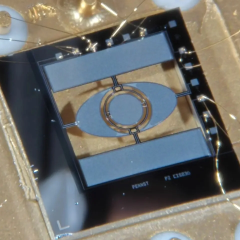
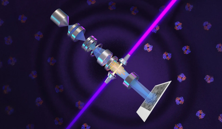
According to НВ — Техно: On October 22 at 22:34, a team of physicists from the University of California, Berkeley announced a breakthrough with a newly developed laser phase plate designed for electron microscopes. This innovation dramatically improves image contrast, enabling researchers to examine tiny proteins and intricate intracellular structures with unprecedented clarity. The advancement marks a significant leap forward in cryo-electron microscopy and tomography, expanding the ability to study molecules as small as 50 kilodaltons, including various proteins.
Phase contrast microscopy was originally pioneered by Fritz Zernike, who won the Nobel Prize in 1953 for this discovery. The new laser-based method manipulates the phase of electron beams to increase image contrast. The design incorporates the world’s most powerful continuous-wave focused laser, confined within a mirrored cavity where the light reflects over 10,000 times, producing high-quality images that were previously unattainable.
Benefits and Future Potential
This cutting-edge technology allows for clear differentiation of small proteins like hemoglobin and key organelles such as mitochondria and nuclei. Cryo-electron tomography (cryo-ET) combines multiple angle images to create detailed 3D models of molecules in their natural cellular environment. While traditional techniques struggled to visualize proteins smaller than 70 kilodaltons, this innovation pushes that boundary down to 50 kilodaltons. Researchers anticipate that future improvements could enable the study of even smaller biological structures, opening new avenues in cellular biology research.
The implications of this development are far-reaching, offering the potential to transform how scientists investigate cells and their components. Enhanced visualization of tiny proteins could deepen our understanding of disease mechanisms and accelerate the creation of new medical treatments. Given the crucial roles proteins play in numerous biological processes, this technology could have a profound impact on both fundamental science and healthcare applications worldwide.
In addition to advancements in microscopy, innovative technologies are also enhancing image analysis in other domains. For example, UCLA has developed a cutting-edge optical-neural processor capable of detecting deepfakes with remarkable accuracy while simultaneously processing multiple video streams. This development underscores the growing intersection of optics and artificial intelligence, further emphasizing the importance of precise imaging techniques across various fields. To learn more about this significant breakthrough, visit UCLA's impressive technology in video analysis.

Read also