

Tracking Mouse Embryo Development Over 13.5 Days Using DNA Typewriter Technology

Developmental Journey
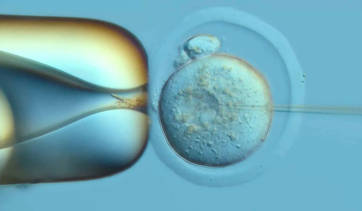
According to НВ — Техно: Researchers at the University of Washington have made a groundbreaking advancement in studying mouse embryo development by employing an innovative tool called DNA Typewriter. This cutting-edge technology enabled scientists to monitor the embryo's growth from the initial division of the fertilized egg through organ formation during the first 13.5 days. DNA Typewriter operates like a biological recording device, embedding a chronological record of cell divisions directly into the DNA.
The study examined 100 fertilized eggs, ultimately yielding 10 embryos suitable for analysis. One embryo was analyzed in exceptional detail, allowing the team to trace nearly all examined cells back to one of the two cells formed after the initial division. Both of these cells contributed nearly equal proportions of various cell types, highlighting their significant roles in subsequent cell specialization.
Significance of the Findings
Scientists discovered that blood cells and retinal cells begin to specialize earlier than the outer skin layer, emphasizing the importance of understanding the precise timing of tissue development, which unfolds at different rates. Previous lineage tracing methods only captured fragments of the cellular ancestry and often involved DNA cutting techniques that risked damaging cells and erasing crucial information.
In contrast, DNA Typewriter records cell division sequences without fully breaking the DNA strand, enhancing reliability for developmental research. This technology holds promise for advancing studies of congenital defects, genetic disorders, cancer, and stem cell biology. While so far tested exclusively on mouse embryos, the method opens new avenues for deciphering cellular biology and organismal development. Considering that mouse pregnancies last 19 to 21 days, insights into these early developmental stages are vital for understanding how organs form.
The discoveries from this research may have profound implications for medical science, particularly in genetics and therapeutic development.
Gaining a clearer picture of how cells develop and specialize early on could pave the way for innovative treatments targeting a range of diseases. Future studies will be essential to validate these findings and expand our knowledge of cellular evolution across different species.
In addition to advancements in embryonic development, recent research has also unveiled a detailed 3D mapping of the mouse brain, highlighting the presence of 539 distinct chemical regions. This innovative approach not only sheds light on white matter but also enhances our understanding of brain structure and function. For insights into how these findings complement the study of cellular development, explore more about the 3D lipid mapping of the mouse brain.

Read also