Laser Plate Boosts Microscopy Resolution by a Factor of 10,000

A Breakthrough in Cryo-Electron Microscopy
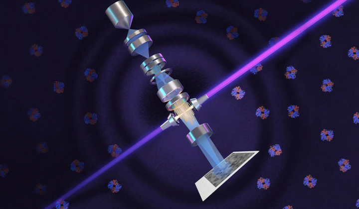
According to НВ — Техно: On June 15 at 9:00 PM, researchers announced the successful adaptation of a phase contrast method for cryo-electron microscopy (cryo-EM). This innovation enables magnifications roughly 10,000 times greater than what conventional light microscopy can achieve. The breakthrough stems from over 15 years of work developing a laser phase plate, which was integrated with the Theia microscope, a system built by Thermo Fisher Scientific.
Testing and Future Prospects
The new system was tested on muscle and blood proteins, with results confirming a substantial improvement in resolution. The instrument is now installed at the University of California, where scientists will continue their research using it. The next step involves implementing cryo-electron tomography (cryo-ET), a technique that combines images taken from multiple angles to create three-dimensional 3D reconstructions.
A powerful laser beam is amplified by highly polished mirrors and focused onto the electron beam to shift its phase. This innovation could have a major impact on research in biology and medicine, opening up new avenues for studying complex molecular structures.
Adapting the phase contrast method for cryo-electron microscopy marks a significant milestone in modern science, as it allows researchers to examine molecules that were previously impossible to observe in such detail. The enhanced resolution could lead to new discoveries in medical and biological research, as well as improvements in diagnostic and treatment methods. The adoption of cryo-electron tomography promises further advancement of this technology, potentially transforming how biological structures are studied at the molecular level.
In light of recent advancements in microscopy, it's noteworthy that scientists have also made significant strides in computational methods. A recent report highlights how researchers have achieved a remarkable 50-fold increase in simulation speed for XFEL experiments. This enhancement not only complements the breakthroughs in cryo-electron microscopy but also promises to accelerate research across various scientific fields.
Read also